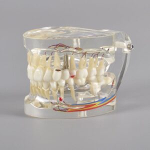
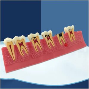
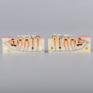
4 X Pathology model with progress of caries
- 4X View Ð Enlarged tooth cross-section for clear visibility of internal structures.
- Caries Progression Ð Shows multiple stages of decay, ideal for explaining dental pathology.
- Detailed & Color-Coded Ð Easy to understand for both students and patients.
- Durable & Lightweight Ð Made from high-quality materials for long-term use.
- Perfect for Clinics & Colleges Ð Enhances communication and learning in professional setups.
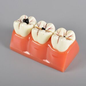
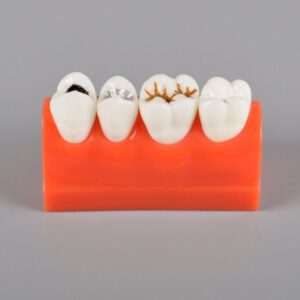
Caries Illustration Model
- Multiple Stages of Decay Ð Clearly shows caries progression from mild to severe.
- Excellent for Comparisons Ð Designed to allow stage-by-stage visual comparisons.
- Patient Education Tool Ð Helps explain the importance of early detection and treatment.
- Durable & Lightweight Ð Made from long-lasting material, easy to handle and carry.
- Ideal for Clinics & Colleges Ð Perfect for demonstrations in dental setups and classrooms.
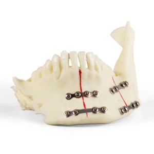
Jaw with titanium plate
- Fracture & Fixation Representation Ð Realistically shows jaw fracture repair with a titanium plate.
- Ideal for Training Ð Designed for surgical demonstrations, dental education, and trauma courses.
- Accurate Anatomical Structure Ð Life-like mandible with visible fracture and metal fixation.
- Durable Construction Ð Built with quality material for long-term classroom or clinical use.
- Patient-Friendly Tool Ð Enhances treatment explanation and boosts patient confidence.
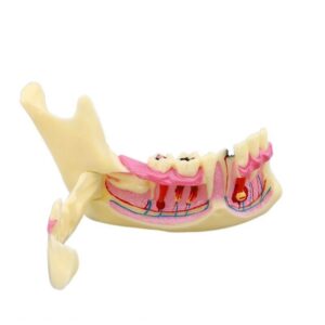
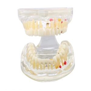
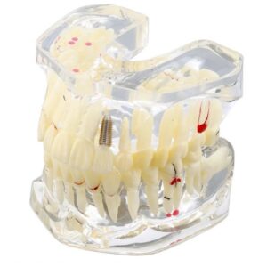
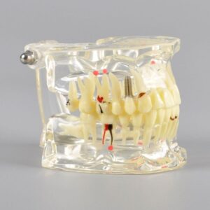
Mandible Model with Caries
- Realistic Caries Depiction Ð Shows common decay patterns on molars and premolars.
- Anatomically Correct Mandible Ð Represents natural jaw structure and tooth positioning.
- Patient-Friendly Tool Ð Simplifies explanation of oral hygiene and cavity treatment.
- Durable and Lightweight Ð Built for repeated use in clinical and academic settings.
- Ideal for Training & Display Ð Useful for dentists, students, and health educators.
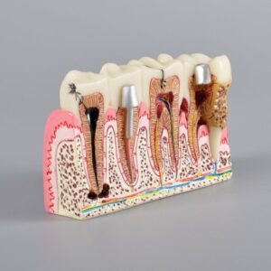
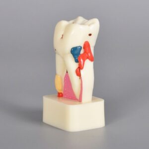
Molar Cross Section Study Model
- Cross-Sectional Detail Ð Displays internal layers: enamel, dentin, pulp, nerve, and vessels.
- Educational Use Ð Ideal for demonstrations in clinics, colleges, and training sessions.
- Durable Construction Ð Made from high-quality, long-lasting material.
- Pathology Ready Ð Highlights conditions like decay or pulp infections clearly.
- Compact & Desk-Friendly Ð Easy to place in study rooms, clinics, or dental classrooms.